Q7:How to confirm the revascularization results?
There are many clinical evaluation methods, the simplest of which is “whether symptoms have improved or not”, which is the most subjective feeling and the most surprising point for participating customers. The next step is the imaging examination. Usually, we recommend that customers go to the hospital to confirm the diagnosis before receiving the vascular reconstruction, keep the complete test report, and then return to the hospital for a second test after the vascular reconstruction is completed to compare with each other. Just compare.
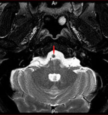
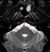
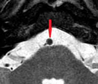
Before Vascular Reconstruction (Computed Tomography):The cross-sectional area of the blood vessel is less than 25% (red arrow)
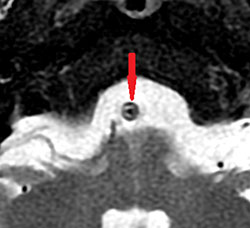
After vascular reconstruction (computed tomography): the vascular cross-sectional area is enlarged to more than 75% (red arrow)(90 days)
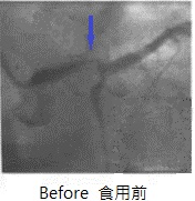
Before revascularization (angiography): blood flow at TIMI 1 (blue arrow)
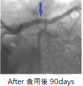
After revascularization (angiography): blood flow at TIMI 3 (blue arrow)
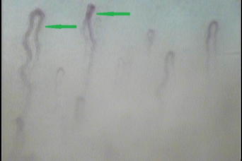
Before revascularization (microvascular photography): 360X (green arrow)
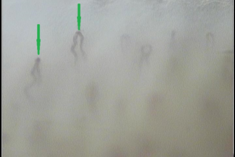
After revascularization (microvascular photography): 360X (green arrow) (40 days)